All posts filed under “3D scanning

comment 0

“Ha! Hold My Brain; Be Still My Beating Heart.”

The post title is a quote from William Mountfort’s play, “Zelmane”, 1705

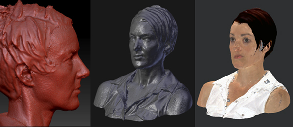
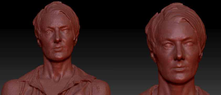
Critical cartographers, like Amy D. Propen suggests that we engage with a map “as something that  both socially constructed and as purporting to represent a “correct” model of the physical world” by understanding cartographic practice as embodied knowledge. This seems especially relevant  when engaging in neuro cartography, or mapping the brain, as this practice – the production of neuroimages – depends on living, embodied, brains.

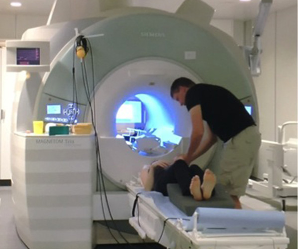
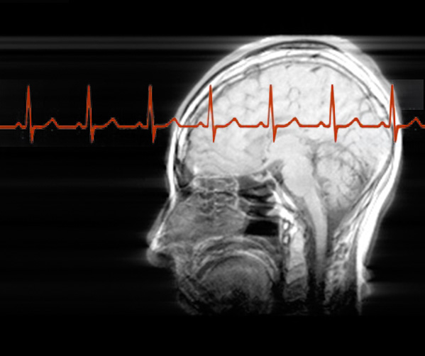
To get the clearest neuroimages my body and brain need to remain very still for the 5-10 minutes that a functional MRI scan takes to complete. This need for stillness makes me self-conscious and that accentuates my sense of being embodied. As I try to control my body and stay still, I  become hyper aware of what are usually unconscious and small bodily movements associated with breathing or swallowing. To move while being scanned adds noise to the data, blurring the image, and therefore the most scientifically useful MRI image emerges through intra-actions that partially erase the trace of my body. ‘Playing dead’ is an essential part of imaging my living brain.

For more see Amy Propen, “Cartographic Representation and the Construction of Lived Worlds: Understanding Cartographic Practice as Embodied Knowledge.” Rethinking Maps. Ed. Martin Dodge, Rob Kitchin, and Chris Perkins. New York: Routledge, 2009. 113-130.

See also, Joseph P. Hornak’s online book, “The Basics of MRI” online.

comment 0

Confessions of a fidget

Since I was a small child I’ve been known as a fidget. So when I was told I had to be very still for a series of seven minute MRI scans I was worried. I was a kid who, when bad dreams sent me running to my parents’ bedroom, kept my mum awake all night as I kicked and wriggled while ‘peacefully’ asleep. That’s the kid that grew into a woman who nightly and tosses and turns. How could I possibly stay still in the scanner? Let along REALLY still. Not even swallowing… Ironically, given the experiments we performed, I needed to still my body, to ‘play dead’ in order to prevent micro movements.

We are usually unaware of how many small movements that we make when we think we are being ‘still’. But in the MRI scanner these micro movements “due to swallowing, fidgeting, overt speech, or transmitted motion as a result of finger pressing on a keypad are a major cause of inconclusive or uninterpretable fMRI results in the clinical setting”. (Desmond et al. 2002). So, my body and brain need to be very still for the 5-10 minutes that a functional MRI scan takes to complete. This stillness created a self-consciousness that accentuated my sense of being embodied. I’d first noticed this during mindfulness meditation, the more mindful I became, the more ‘bodiful’ I realized I was. As I quieten my mind, my awareness is more open to, and often overwhelmed by the sensations in my body. Little aches and pains, the feels of the surface I am lying on, the temperature of the air as it fills my lungs, all these sensations flood in. As I practiced, knowing I would have to lie very still and not even swallow in the scanner, I found myself trying to control what are usually unconscious and small bodily movements, like those associated with breathing or swallowing. As a newbie mindfulness practitioner and a wannabe MRI subject I became hyper aware of my tiny movements and that awareness became tension which resulted in ticks and twitches. My ongoing lesson is to have the awareness but not to focus on it, not to turn it into tension. I noticed in the moment of imagining resisting movement, I would make a small movement. It was a sort of mental ‘tensing’ as I thought of wanting to move, and then tried to stop that thought, right then my muscles would alter as I imagined ‘not moving’.

This project is full of surprises, it’s made me know more about myself and taught me new things. But one of the biggest surprises was when I came out of the MRI scanner and the neuroscientist, Torben, commented on how still I’d been. I was stunned (and proud of myself). After 50 years of fidgeting and being told off for it, I am learning to stay still!

Desmond, John E, and Annabel S. H. Chen. “Ethical issues in the clinical application of fMRI: factors affecting the validity and interpretation of activations.” Brain and cognition  50, no. 3 (2002): 482-497.

comment 0

Self portrait #1

The first neuro memento mori self portrait we created combines the structural MRI of my brain with the structural 3D scans of my face, head and neck. This piece does not show brain activity but is formally similar to memento mori and vanitas works. However, it takes memento mori a step further by not only showing the skull but cutting into the skull to show the artist’s brain. The object is life-sized. It is displayed at the new Moesgaard Museum, Aarhus, Denmark as part of the ethnographic exhibition, ‘The Life of the Dead’ which runs 2014-2018.

comment 0

I’m melting, I’m melting…

Sometimes you don’t get the data that you were expecting… When we transferred data between programs we had a 3D model like this. It reminded me of the caul found occasionally on newborn babies. The name caul or cowl (Latin: Caput galeatum, literally, “helmeted head”) is a piece of membrane that can cover a newborn’s head and face. It’s a little ghostly, somehow appropriate to a memento mori project, like one of the vanitas memento mori waxworks melting, or my face as if covered in a muslin death shroud. I’m thinking of 3D printing it as it is. It’s a happy accident, serendipity.

comment 0

Head scanning and hair gel

599-jane-prophet-neuro-memento-mori-3D-face-scan01
We tried a couple of different scanners for the head scan to make the 3D model of my face and neck. Shadow and dark hair is hard to scan and the top photo shows how the technician passes the scanner with a bright light under my chin to scan the shadowy area. Many people with black hair dust their hair with a white talcum powder to make it easier for the scanner to ‘see’. The fine strands of hair are also hard to capture – this is like the early days of green screen technology in video with hair looking fuzzy. My solution was to work with that constraint and so I hair gelled my hair to give it more shape and definition which was easier for the scanner to pick up. I kind of like the Max Headroom look!

We used two hand-held 3D scanners, first the Go!SCAN scanner which captured high detail and facial geometry data. As a single scan covers a limited surface area, moving the scanner to make multiple scans is necessary to create a complete a facial model that is accurate and detailed. We had captured the rough shape of the heavily gelled hair, but the Go!SCAN’s high resolution struggled to identify the detail of the hair structure so we switched to another scanner. The Sense scanner by Cubify scans at lower definition but suited our purpose – to capture the hair as a larger solid shape.

jane-prophet-neuro-memento-mori-3D-face-scan02
The result is this model. Some parts of the left hand side image, such as the mouth, look distorted. We think that this is because it is difficult for a person to keep perfectly still during the multiple passes of the handheld scanner and Jane smiled part way through. Every subtle movement leads to displacement and distortion in the final results. The image on the right shows the model after the distortions were fixed, fairly quickly, by Ivan using ZBrush sculpting tools  and photo references. Finally, the model was completely remeshed to redistribute vertices evenly and to reduce the vertices count and close holes, making the model 3D-printing friendly.